상품상세정보
의학서적전문 "성보의학서적"의 신간의학도서입니다.
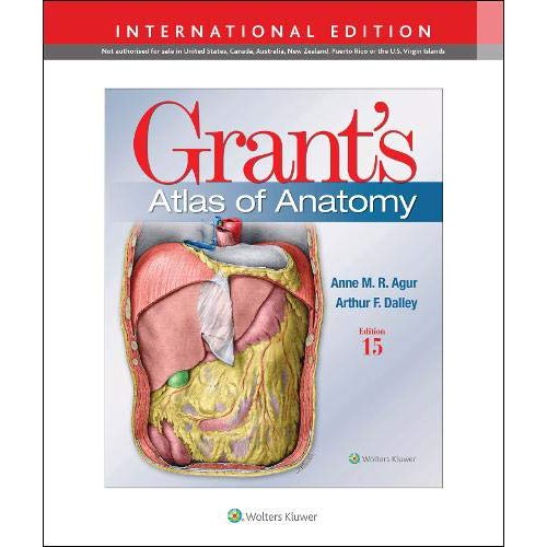
For more than seventy-five years, Grant’s Atlas of Anatomy has maintained a tradition of excellence while continually adapting to meet the needs of each generation of students. The updated fifteenth edition is a visually stunning reference that delivers the accuracy, pedagogy, and clinical relevance expected of this classic atlas, with new and enhanced features that make it even more practical and user-friendly.
Illustrations drawn from real specimens, presented in surface-to-deep dissection sequence, set Grant’s Atlas of Anatomy apart as the most accurate reference available for learning human anatomy. These realistic representations bring structures to life and provide students with the ultimate lab resource.
• NEW Illustration overviews highlight the autonomic nerves to clarify nerve and muscle innervations.
• NEW and updated images reflect the latest clinical insights through:
• 15 NEW Illustrations
• 160 revised figures
• NEW and updated surface anatomy images
• NEW pulses photos
• Renowned, high-resolution, dynamically colored illustrations organized in dissection sequence enable the formation of 3-D constructs for each body region and provide detailed, realistic reference during dissection.
• Tables detail muscles, vessels, and other anatomic information in an easy-to-use format ideal for review and study.
• Enhanced medical imaging includes more than 100 clinically significant MRIs, CT images, ultrasound scans, and corresponding orientation drawings to help students confidently apply the laboratory experience to clinical rotations.
• Color schematic illustrations reinforce the relationships of structures and anatomical concepts in vibrant detail.
- Table of Contents -
Pectoral Region 88
Axilla, Axillary Vessels, and Brachial Plexus 95
Scapular Region and Super? cial Back 106
Arm and Rotator Cuff 110
Joints of Shoulder Region 124
Elbow Region 132
Elbow Joint 138
Anterior Forearm 144
Anterior Wrist and Palm of Hand 152
Posterior Forearm 168
Posterior Wrist and Dorsum of Hand 171
Lateral Wrist and Hand 176
Medial Wrist and Hand 179
Bones and Joints of Wrist and Hand 180
Function of Hand: Grips and Pinches 186
Imaging and Sectional Anatomy 187
CHAPTER 1
BACK ............................................................... 1
Overview of Vertebral Column 2
Cervical Spine 8
Craniovertebral Joints 12
Thoracic Spine 14
Lumbar Spine 16
Ligaments and Intervertebral Discs 18
Bones, Joints, and Ligaments of Pelvic Girdle 23
Anomalies of Vertebrae 29
Surface Anatomy of Back 30
Muscles of Back 33
Suboccipital Region 42
Spinal Cord and Meninges 44
Vertebral Venous Plexuses 52
Components of Spinal Nerves 53
Dermatomes and Myotomes 56
Autonomic Nerves 58
Imaging of Vertebral Column 62
CHAPTER 2
UPPER LIMB ................................................... 65
Systemic Overview of Upper Limb: Bones 66
Systemic Overview of Upper Limb: Nerves 72
Systemic Overview of Upper Limb: Arteries 80
Systemic Overview of Upper Limb: Veins and Lymphatics 82
Systemic Overview of Upper Limb: Musculofascial
Compartments 86xii Contents
CHAPTER 3
THORAX ...................................................... 193
Pectoral Region 194
Breast 196
Bony Thorax and Joints 204
Thoracic Wall 211
Thoracic Contents 219
Pleural Cavities 222
Mediastinum 223
Lungs and Pleura 224
Bronchi and Bronchopulmonary Segments 230
Innervation and Lymphatic Drainage of Lungs 236
External Heart 238
Coronary Vessels 250
Conduction System of Heart 254
Internal Heart and Valves 255
Superior Mediastinum and Great Vessels 262
Diaphragm 269
Posterior Thorax 270
Overview of Autonomic Innervation 280
Overview of Lymphatic Drainage of Thorax 282
Sectional Anatomy and Imaging 284
Dr. John Charles Boileau Grant vi
Reviewers vii
Preface viii
Recoloring Grant’s Atlas ix
Acknowledgments x
List of Tables xiv
Figure and Table Credits xvi
References xix
CHAPTER 4
ABDOMEN .................................................. 291
Overview 292
Anterolateral Abdominal Wall 294
Inguinal Region 304
Testis 314
Peritoneum and Peritoneal Cavity 316
Digestive System 326
Stomach 327
Pancreas, Duodenum, and Spleen 330
Intestines 334
Liver and Gallbladder 344
Biliary Ducts 354
Portal Venous System 358
Posterior Abdominal Viscera 360
Kidneys 363
Posterolateral Abdominal Wall 367
Diaphragm 372
Abdominal Aorta and Inferior Vena Cava 373
Autonomic Innervation 374
Lymphatic Drainage 380
Sectional Anatomy and Imaging 384
CHAPTER 5
PELVIS AND PERINEUM ................................ 391
Pelvic Girdle 392
Ligaments of Pelvic Girdle 399
Floor and Walls of Pelvis 400
Sacral and Coccygeal Plexuses 404
Peritoneal Re? ections in Pelvis 406
Rectum and Anal Canal 408
Organs of Male Pelvis 414
Vessels of Male Pelvis 420
Lymphatic Drainage of Male Pelvis and Perineum 422
Innervation of Male Pelvic Organs 424
Organs of Female Pelvis 426
Vessels of Female Pelvis 436
Lymphatic Drainage of Female Pelvis and Perineum 438
Innervation of Female Pelvic Organs 440
Subperitoneal Region of Pelvis 444
Surface Anatomy of Perineum 446
Overview of Male and Female Perineum 448
Male Perineum 453
Imaging of Male Pelvis and Perineum 460
Female Perineum 462
Imaging of Female Pelvis and Perineum 468
Pelvic Angiography 470
CHAPTER 6
LOWER LIMB ............................................... 471
Systemic Overview of Lower Limb: Bones 472
Systemic Overview of Lower Limb: Nerves 476
Systemic Overview of Lower Limb: Blood Vessels 484
Systemic Overview of Lower Limb: Lymphatics 488
Systemic Overview of Lower Limb: Musculofascial
Compartments 490
Retro-inguinal Passage and Femoral Triangle 492
Anterior and Medial Compartments of Thigh 496
Lateral Thigh 503
Bones and Muscle Attachments of Thigh 504
Gluteal Region and Posterior Compartment of Thigh 506
Hip Joint 516
Knee Region 522
Knee Joint 528
Anterior and Lateral Compartments of Leg,
Dorsum of Foot 542
Posterior Compartment of Leg 552
Tibio? bular Joints 562
Sole of Foot 563
Ankle, Subtalar, and Foot Joints 568
Imaging and Sectional Anatomy 581
CHAPTER 7
HEAD ........................................................... 585
Cranium 586
Face and Scalp 606
Meninges and Meningeal Spaces 615
Cranial Base and Cranial Nerves 620
Blood Supply of Brain 626
Orbit and Eyeball 630
Parotid Region 642
Temporal Region and Infratemporal Fossa 644
Temporomandibular Joint 652
Tongue 656
Palate 662
Teeth 665
Nose, Paranasal Sinuses, and Pterygopalatine Fossa 670
Ear 683
Lymphatic Drainage of Head 696
Autonomic Innervation of Head 697
Imaging of Head 698
Neuroanatomy: Overview and Ventricular System 702
Telencephalon (Cerebrum) and Diencephalon 705
Brainstem and Cerebellum 714
Imaging of Brain 720Contents xiii
CHAPTER 8
NECK ........................................................... 725
Subcutaneous Structures and Cervical Fascia 726
Skeleton of Neck 730
Regions of Neck 732
Lateral Region (Posterior Triangle) of Neck 734
Anterior Region (Anterior Triangle) of Neck 738
Neurovascular Structures of Neck 742
Visceral Compartment of Neck 748
Root and Prevertebral Region of Neck 752
Submandibular Region and Floor of Mouth 758
Pharynx 762
Isthmus of Fauces 768
Larynx 774
Sectional Anatomy and Imaging of Neck 782
CHAPTER 9
CRANIAL NERVES ......................................... 787
Overview of Cranial Nerves 788
Cranial Nerve Nuclei 792
Cranial Nerve I: Olfactory 794
Cranial Nerve II: Optic 795
Cranial Nerves III, IV, and VI: Oculomotor, Trochlear,
and Abducent 797
Cranial Nerve V: Trigeminal 800
Cranial Nerve VII: Facial 806
Cranial Nerve VIII: Vestibulocochlear 808
Cranial Nerve IX: Glossopharyngeal 810
Cranial Nerve X: Vagus 812
Cranial Nerve XI: Spinal Accessory 814
Cranial Nerve XII: Hypoglossal 815
Summary of Autonomic Ganglia of Head 816
Summary of Cranial Nerve Lesions 817
Imaging of Cranial Nerves 818
Index 821
기타 도서와 관련된 문의사항은 고객센터(02-854-2738) 또는 저희 성보의학서적 홈페이지내 도서문의 게시판에 문의바랍니다.
감사합니다.
성보의학서적 "http://www.medcore.kr


